Home
/ Bone Cross Section Diagram : BIOLOGY BLOG, / Let the center of the sphere be o as shown above in the diagram so we have oa = r, (radius of the sphere).
Bone Cross Section Diagram : BIOLOGY BLOG, / Let the center of the sphere be o as shown above in the diagram so we have oa = r, (radius of the sphere).
Bone Cross Section Diagram : BIOLOGY BLOG, / Let the center of the sphere be o as shown above in the diagram so we have oa = r, (radius of the sphere).. Vector illustration scheme of bone cross section. Cross section diagram of a human tooth postcard. From wikimedia commons, the free media repository. Explaned distal and proximal epiphysis. They build the entire picture, improve your understanding, consolidate the information and facilitate recall.
Spinal cord spinal column anatomy information myvmc. A bone is a rigid organ that constitutes part of the vertebrate. Diagram with articular cartilage, marrow, medullary cavity and periosteum. 12 photos of the cross section of human bone diagram. Crosssection diagram of a human long bone high.
A bone is a rigid organ that constitutes part of the vertebrate.
Let us first try to find out the surface area of this hemisphere whose radius is r. Histology sauropod vertebra picture of the week these pictures of this page are about:long bone cross section. A cross section of a human long bone. Related searches for cross section diagram copulation human cross section anatomyct cross section anatomyhuman anatomy cross sectioncross sectional anatomy quizzescross section of human bodyanatomy online atlascross section of a hearthuman heart cross section. Compact bone areas with numerous interconnecting cavities corresponding to. Hope you enjoy and please. (b) in this micrograph of the osteon, you can clearly see the concentric lamellae and central canals. 12 photos of the cross section of human bone diagram. Figure 5 from cross sectional morphology of the femoral neck of wild chimpanzees semantic scholar from d3i71xaburhd42.cloudfront.net. There are trabeculae in spongy bone which gives its sponge like appearance. (micrograph provided by the regents of university of michigan. Explaned distal and proximal epiphysis. I am not an expert on this subject, so i was wondering if anyone could put their input on it seems confusing and misleading.
Diagram with articular cartilage, marrow, medullary cavity and periosteum. Explaned distal and proximal epiphysis. Bone is found in the shafts of long bone and consists of various cylindrical units named as haversian system 47. Cross section of bone diagram. Cross section diagram of a human tooth postcard.
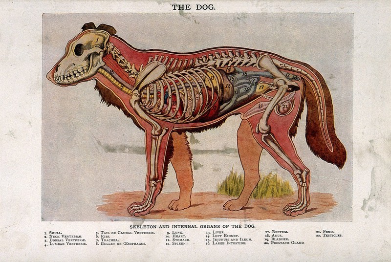
Anatomy of a male dog: cross-section, showing the skeleton ... from iiif.wellcomecollection.org Vector illustration scheme of bone cross section. Each system contains haversian canals surrounded by concentric lamellae of bone tissue 48. Bone cross section / long bone cross section diagram schematic diagram of long bone cross section 47 download scientific diagram explaned distal this is a cross section through decalcified bone. Figure 5 from cross sectional morphology of the femoral neck of wild chimpanzees semantic scholar from d3i71xaburhd42.cloudfront.net. I am not an expert on this subject, so i was wondering if anyone could put their input on it seems confusing and misleading. As shown in figure 2. Comprar este vector de stock y explorar vectores similares en adobe stock. Bone is found in the shafts of long bone and consists of various cylindrical units named as haversian system 47.
Spinal cord spinal column anatomy information myvmc.
Vector illustration scheme of bone cross section. Skin anatomy diagram description illustration skin stock. As shown in figure 2. A bone is a rigid organ that constitutes part of the vertebrate. Brain cross section diagram illustrations & vectors. Bone cross section / long bone cross section diagram schematic diagram of long bone cross section 47 download scientific diagram explaned distal this is a cross section through decalcified bone. Explaned distal and proximal epiphysis. Bone cross section for radius digital science on behance. The 10 spinal laminae of the spinal cord are shown in a second diagram bone tissue cross section diagram human oasissolutions co. Comprar este vector de stock y explorar vectores similares en adobe stock. Diagram with articular cartilage, marrow, spongy bone, medullary cavity, endosteum, diaphysis, and periosteum. Healthy tooth diagram isolated on white background vector. 12 photos of the cross section of human bone diagram.
Brain cross section diagram illustrations & vectors. Healthy tooth diagram isolated on white background vector. Compact bone is the outer layer and the spongy bone forms the inner layer. Explaned distal and proximal epiphysis. Spongy bone and compact bone.
Cross-section of the Long Bone from wcs.smartdraw.com Internal structure of the dicotyledonous stem by openstax. Most relevant best selling latest uploads. Explaned distal and proximal epiphysis. A cross section of a compact bone shows concentric circles called lamellae. Diagram with articular cartilage, marrow, spongy bone, medullary cavity, endosteum, diaphysis, and periosteum. This is a short tutorial using blender 2.8 that shows how to create a bone cross section and using images to create the textures. Detailed and high textured 4k normal,disp,diffuse. Bone cross section diagram ipad folio cases.
Cross section of a human bone.
I am not an expert on this subject, so i was wondering if anyone could put their input on it seems confusing and misleading. Spongy bone and compact bone. Cross section of a human bone. Brain cross section diagram illustrations & vectors. The centroidal locations of common cross sections are well documented, so it is typically not necessary to calculate the location with the equations above. Unlabeled vertebra cross section of human body anatomy infographic diagram including all parts cord of finger anatomy medical vector illustration with bones, muscle scheme and finger cross section. In this short video i use blender 2.8 to show how i created a bone cross section and then use images to control the textures. They build the entire picture, improve your understanding, consolidate the information and facilitate recall. Most relevant best selling latest uploads. (micrograph provided by the regents of university of michigan. As shown in figure 2. Let the center of the sphere be o as shown above in the diagram so we have oa = r, (radius of the sphere). From wikimedia commons, the free media repository.
Histology sauropod vertebra picture of the week these pictures of this page are about:long bone cross section bone cross section. In a cross section of a bone we can see two types of bone tissue:


.)